Author: Hajira Mahmood
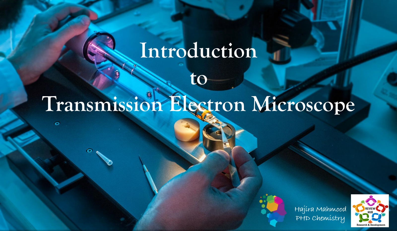
A transmission electron microscope (TEM) is a powerful tool used to observe the ultrastructure of materials at a very high resolution. It uses a beam of electrons to create an image of the specimen.

Basic Principles of Transmission Electron Microscope
The fundamental principle of a TEM involves an electron beam passing through an ultrathin specimen. This creates an image by interacting with the specimen and forming a magnified projection. The microscope operates based on the wave-like properties of electrons and the resulting interactions with the specimen surface.
Components of a Transmission Electron Microscope

Electron Gun
The electron gun generates a beam of electrons that will be used to scan the specimen, facilitating imaging and analysis.
Electromagnetic Lenses
These lenses focus and control the path of the electron beam in order to produce a clear image of the specimen.
Detectors
Detectors capture and process the electron signals, which are then transformed into a visual representation of the specimen.
Operating Procedure of a Transmission Electron Microscope
The operating procedure of a TEM involves various steps, including sample preparation, setting the appropriate voltage, aligning the lenses, and adjusting the focus.
Sample Preparation
Specimens need to be ultra-thin and properly mounted on a grid to be compatible with the TEM imaging process.
Voltage Adjustment
Setting the correct voltage ensures the electron beam can penetrate the specimen to produce a clear image.
Focusing and Imaging
Aligning, focusing, and capturing images of the specimen is the final phase of the operating procedure.
Applications of Transmission Electron Microscope
Material Science
TEM is crucial for analyzing the microstructure and composition of materials at the atomic and molecular levels.
Life Sciences
TEM is used for investigating biological samples, such as cells, tissues, and viruses, at high resolution.
Nanotechnology
TEM plays a significant role in visualizing and characterizing nanomaterials and nanostructures.
Pharmaceuticals
TEM is employed in pharmaceutical research to study drug formulations and microstructures of materials.
Advantages of Using a Transmission Electron Microscope
High Resolution Imaging
TEM provides exceptionally high-resolution images, enabling the visualization of structures at the atomic scale.
Elemental Analysis
It allows for elemental analysis of samples using techniques like energy-dispersive X-ray spectroscopy (EDS).
Nanometer Scale Observation
TEM can observe and analyze features at the nanometer scale, offering insights into nanomaterials and devices.
Limitations of a Transmission Electron Microscope
Sample Preparation Challenges
Preparing samples for TEM requires meticulous techniques, often leading to complex and time-consuming procedures.
Specimen Damage
The high-energy electron beam can cause damage to certain specimens, affecting the accuracy of the results.
Operational Expertise
Operating and understanding the complex instrumentation of TEM demands specialized skills and training.
Conclusion and Future Developments in Transmission Electron Microscopy
Transmission electron microscopy continues to advance, with ongoing developments aimed at enhancing imaging resolution, improving ease of use, and expanding its applications in various fields.
The future holds promise for TEM innovations that could unlock new frontiers in nanotechnology, materials science, and life sciences.
Also read: Mastering the Art of Chemical Synthesis: Unveiling Methods and Marvels
Follow Us on

